Overview of Immunodiagnosis
(2022年12月27日)https://www.sekbio.com/services/
1. Basic concept
(1) Antibodies (Ab) and antigens (Ag)
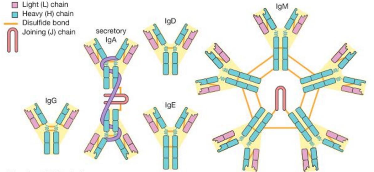
Antibody: It refers to the protective protein produced by the body due to antigen stimulation, secreted by effector B cells in vertebrates, which can be divided into IgG, IgM, IgA, IgD, and IgE. IgG is the isotype with the highest content in serum and body fluids, accounting for 75% to 80%. IgG is the primary antibody produced by the secondary immune response, the "main force" of the body's anti-infection, and the most used antibody in immunodiagnosis.
Antigen: It refers to a substance that can cause the production of antibodies and any substance that can induce an immune response in the body. Antigens are foreign, macromolecular, and specific. The specificity of an antigen means that an antigen can only bind to the corresponding antibody or effector T cells, which chemical groups determine on the molecule's surface, called antigenic determinants. The molecular weight of the antigen is generally >10kDa, and the larger the molecular weight, the stronger the antigenicity.
(2) Polyclonal antibodies and monoclonal antibodies
Polyclonal antibody: natural antigen molecules often contain a variety of different antigenic epitopes, stimulating the body's immune system with this antigen can activate a variety of B cell clones at the same time, and the resulting antibodies will contain a variety of antibodies against different epitopes, so they are called polyclonal antibodies.
The monoclonal antibody is a highly homogeneous antibody produced by a single B cell clone that only targets a specific epitope. It is typically prepared using hybridoma technology. Effector B cells and myeloma cells are fused to form B cell hybridomas. A monoclonal antibody can produce by culturing a single hybridoma cell into a population of cells specific for an epitope. The affinity and specificity of monoclonal antibodies are generally better than those of polyclonal antibodies.
(3) Recombinant antibodies and recombinant antigens
Recombinant antibody: A genetically engineered antibody is an antibody produced in vitro using gene recombination technology and protein engineering technology. Obtain the gene sequence of the antibody, construct an expression vector, and transfer it into an expression host (such as yeast, insect cells, mammalian cells, or bacteria). In this way, the production of antibodies is freed from the constraints of the immune system. At the same time, both full-length antibodies and various antibody fragments can produce.
Recombinant antigens: Generally, the recombinant antigens refer to recombinant proteins in essence and are also proteins produced in vitro by gene recombination technology and protein engineering technology.
2. Principles of immunodiagnosis
(1) Method of double monoclonal antibody sandwich
Double monoclonal antibody sandwich method: use a solid phase carrier to coat one antibody and label another labeled antibody to detect the antigen to be tested, and then form a complex of coated Ab-Ag-labeled Ab. This method mainly detects antigenic substances.
Image credit:https://www.rockland.com/resources/elisa-technique/
(2) Indirect method
Indirect method: a detection method in which a solid-phase carrier is used to immobilize the antigen and label the secondary antibody. This method is mainly used to detect IgG antibodies in samples. A complex of the coated Ag-target Ab-labeled secondary Ab is formed to detect the antibody in the sample.
Image credit:https://www.rockland.com/resources/elisa-technique/
(3) Capture method
Capture method: a detection method in which a solid-phase carrier is used to coat the secondary antibody and another strain of labeled antigen to detect the antibody. A complex of the coated secondary Ab-target Ab-labeled Ag is formed and tested. This method is mainly used to detect IgM in the sample.
Image credit:https://www.rockland.com/resources/elisa-technique/
(4) Competition method
Competition method: Competitive binding of the coated/labeled antibody with the labeled/coated antigen and the test antigen. This method is mainly used to detect minor molecule antigens.
Image credit:https://www.rockland.com/resources/elisa-technique/
Method 1: coat the antibody on the solid phase, label the antigen, add the sample, the antigen to be tested competes for the antibody binding site, wash, output the result
Method 2: coat the antigen on the solid phase, label the antibody
3. The development of immunodiagnosis
Immunodiagnosis applies immunology theories, techniques, and means to diagnose various diseases and determine immune status. With technology development, immunodiagnosis has undergone immunoelectrophoresis, precipitation and agglutination assays, radioimmunoassay, immunofluorescence, enzyme-linked immunosorbent assay, immunoturbidimetry, colloidal gold and fluorescent microsphere labeling, blotting membrane strips, chemiluminescence, protein chips, microfluidics control technology and single-molecule detection.
(1) Enzyme-linked immunosorbent assay (ELISA)
Enzyme-linked immunosorbent assay (ELISA) is a detection method that combines the immune reaction principle with immobilization technology and enzyme labeling technology. First, use a polystyrene multi-well plate to coat the antigen/antibody, bind the substance to be tested in the sample, and add horseradish peroxidase (HRP) or alkaline phosphatase (AP) after washing to label the antibody/antigen to form an immune complex. Then add an enzymatic chromogenic substrate and a stop solution to terminate the reaction. Finally, qualitatively or quantitatively determine the sample content analyte by the color OD.
Image credit:https://www.biotekchina.com.cn/applications/elisa-and-related-immunoassays.html
(2) Colloidal gold/fluorescence immunochromatography
Colloidal gold: refer to the gold sol with the dispersed phase particle diameter between 1 and 150 nm, and the color is orange-red to purple-red.
Colloidal gold immunochromatography: a detection technique that uses colloidal gold-labeled antibody, use nitrocellulose membrane (NC membrane) as a solid phase carrier to coat antigen/antibody, and uses the capillary action of the NC membrane to chromatograph the liquid from one end of the membrane to the other end, at the same time, an immune reaction occurs during the chromatography.
Fluorescence immunochromatography: an immunochromatographic detection technology alters the colloidal gold into fluorescent microsphere labeling.
(3) Chemiluminescence immunoassay
Chemiluminescence: refers to the emission of light that accompanies the process of chemical reactions.
Chemiluminescence immunoassay method: a detection technique that uses multi-well plate or magnetic particles as solid carriers to coat antigen/antibody, add analyte and luminophore to label antigen/antibody to form an immune complex, after washing, chemically stimulate the complex or substrate to emit light, and determines the content of the analyte by the luminescence intensity.
Image credit:https://www.sepmag.eu/blog/bid/335317/the-two-key-reasons-for-selecting-chemiluminescence-for-immunoassays
(4) Classification of chemiluminescence immunoassay
Classification by coating carrier: It is divided into plate chemiluminescence and magnetic particle chemiluminescence. At present, plate chemiluminescence has been eliminated.
Classification by luminescence: divided into indirect luminescence (enzymatic), direct luminescence (acridine ester, luminol), and electrochemiluminescence.
Sekbio provides chemi luminescent immuno assay, chemiluminescent assay, chemiluminescence immunoassay, etc. For more information, please feel free to contact us!
- このできごとのURL:


コメント