TOF 3D imaging boosts surgical precision and real-time medical diagnosis
(2025年10月27日)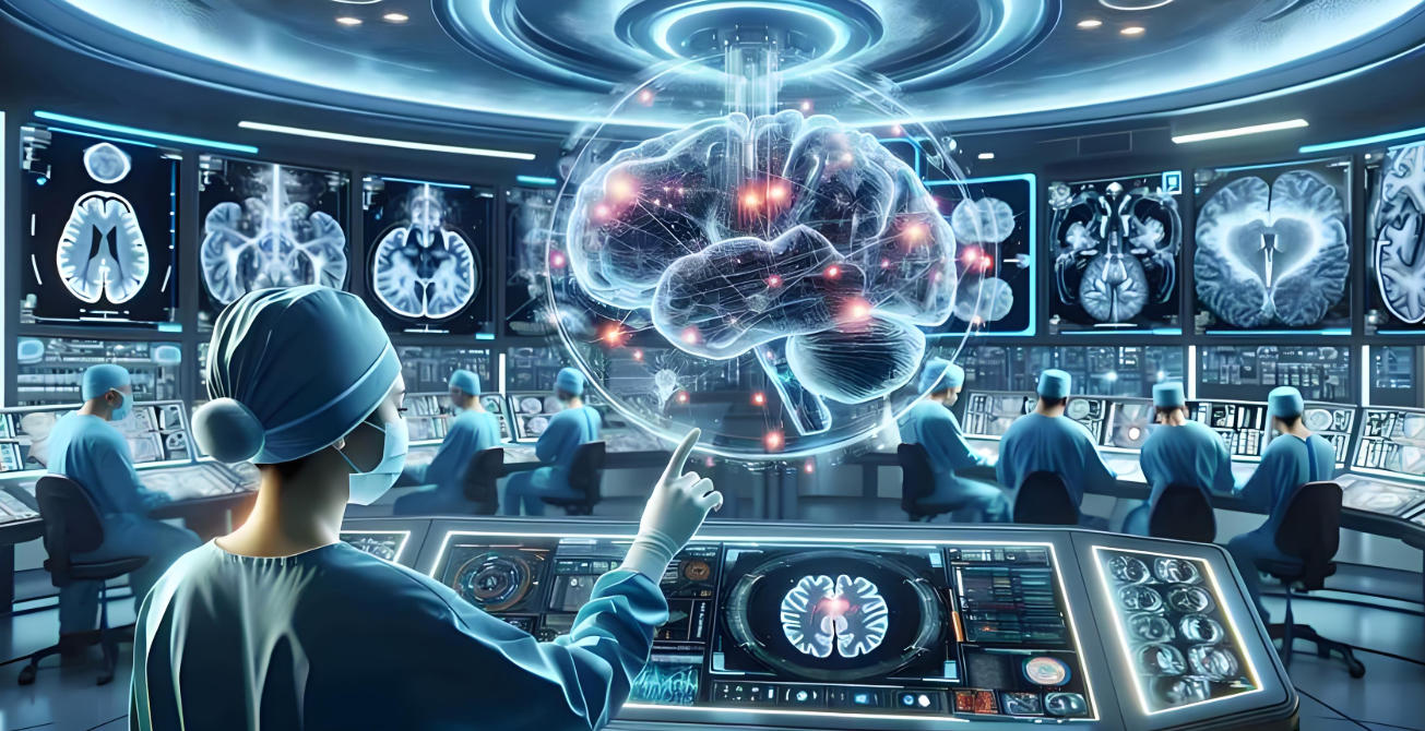
How TOF Technology Enables Precise Diagnosis and 3D Medical Imaging in Minimally Invasive Surgeries
What is Time-of-Flight (TOF) Depth Sensing Technology for Medical Imaging?
Time-of-Flight (TOF) technology is a depth-sensing method based on measuring the travel time of light (usually infrared pulses) as it travels from the emitter to a target and back to the sensor. In medical imaging, TOF systems send out light pulses or modulated waves, then measure the time difference or phase shift of reflections to calculate precise distances and generate 3D spatial maps.
In medical diagnostics and surgical imaging, TOF offers several distinct advantages:
3D Depth Perception: Provides per-pixel depth data for reconstructing organs, lesions, and tissue surfaces in three dimensions.
Non-contact Measurement: Allows non-invasive and minimally invasive operations by collecting data via reflected light rather than direct contact.
High Real-time Performance: Delivers millisecond-level feedback, supporting real-time surgical navigation and dynamic tissue monitoring.
Multi-scenario Applicability: Suitable for surgical planning, intraoperative navigation, rehabilitation monitoring, and intelligent healthcare systems.
By transforming time measurements into 3D depth data, TOF brings real-time, radiation-free, and high-precision imaging capabilities to modern medicine.
Limitations of Traditional Medical Imaging & Why 3D TOF Technology Matters
Traditional medical imaging—such as X-ray, CT, MRI, and 2D ultrasound—remains the diagnostic backbone but struggles to meet the requirements of minimally invasive and precision surgeries.

Key Limitations
Lack of 3D Spatial Information
2D images provide only flat projections. They fail to reflect the true spatial structure and depth of organs or lesions, making it difficult to plan accurate incisions or avoid vital tissues.
Inaccurate Surgical Planning
Minimally invasive surgeries require precise paths through limited spaces. Planning based on 2D images often involves guesswork about depth and spatial relationships, increasing the risk of surgical deviation or instrument misplacement.
Low Diagnostic Efficiency
2D imaging requires multiple slices and high operator experience to interpret spatial relationships, which may slow diagnosis and increase error rates.
Insufficient Real-time Tracking
Organs move due to breathing or heartbeat. Traditional methods lack real-time feedback, making it difficult to maintain accuracy during dynamic surgical procedures.
Why TOF 3D Imaging Is the Solution
TOF imaging overcomes these issues by providing:
Real-time volumetric visualization instead of flat 2D projections.
Dynamic 3D tracking for moving organs and instruments.
Precise volumetric measurement of lesions and tissue structures.
Improved preoperative planning and intraoperative navigation accuracy.
How TOF Technology Facilitates Precise Diagnosis and 3D Medical Imaging in Minimally Invasive Surgeries
1. Real-time Depth Scanning
TOF sensors emit modulated light pulses and measure reflection time to construct 3D maps within milliseconds. Surgeons can visualize organs, tools, and lesions in real time, making adjustments instantly during surgery.
2. Accurate Volume Measurement
TOF imaging creates full 3D point clouds, allowing precise measurement of organ and lesion volume. Surgeons can calculate resection boundaries more accurately, minimizing collateral tissue damage.

3. Precise Lesion Localization
By generating true 3D spatial coordinates, TOF imaging pinpoints lesion locations and nearby critical structures, guiding surgeons in planning minimally invasive paths and avoiding damage to vital anatomy.
4. Dynamic Tissue Tracking and Navigation
TOF sensors continuously track tissue movement caused by breathing or heartbeats. When integrated with robotic systems or navigation software, this enables surgeons to adjust movements dynamically and maintain precision.
5. AI-Assisted Diagnosis and Personalized Treatment
With AI integration, TOF 3D data supports lesion recognition, risk prediction, and surgical simulation. Patient-specific 3D anatomical modeling enables personalized surgery and rehabilitation planning.
Clinical Applications of TOF in Medical Imaging
a) Minimally Invasive Surgical Navigation
TOF-based 3D imaging provides high-accuracy navigation for laparoscopic, neurosurgical, and robotic operations. Surgeons can plan optimal incision points, minimize tissue disruption, and adjust intraoperative strategies dynamically.
b) Tumor Localization and Volume Measurement
TOF scans help identify tumor boundaries and calculate exact tumor volumes. Real-time monitoring enables adaptive resection and supports precise radiotherapy or drug delivery.
c) Vascular Imaging and Interventional Guidance
3D TOF imaging allows accurate visualization of blood vessel structures for catheter and stent placement. It minimizes the risk of vascular injury and enhances procedural control during interventional treatments.
d) Personalized Rehabilitation and Monitoring
TOF enables post-operative tracking of joint movement, gait, and recovery progress. Patients can undergo contact-free monitoring, and data can be used for remote medical consultation and AI-based rehabilitation evaluation.
Technical Challenges and Optimization Strategies
Despite its advantages, TOF still faces practical challenges in clinical applications, mainly due to soft tissue reflectivity, scattering, and motion interference.
Technical Challenges
Low Reflectivity in Soft Tissues: Biological tissues attenuate infrared light, leading to signal loss and depth noise.
Light Scattering and Multipath Effects: Scattering causes inaccuracies in depth data and reduced spatial resolution.
Organ Motion Interference: Respiratory and cardiac motion cause instability in real-time depth measurements.
Resolution and Field-of-View Limits: Some sensors lack sufficient precision for delicate surgical procedures.

Optimization Approaches
Optimized Wavelengths: Use near-infrared light for better penetration and stable reflections.
AI-Enhanced Signal Processing: Apply deep learning to denoise, correct deviations, and stabilize real-time imaging.
Multi-Sensor Fusion: Combine TOF with CT, MRI, or ultrasound for complementary imaging that merges real-time depth with high-resolution detail.
Hardware Upgrades: Develop low-noise, high-sensitivity TOF sensors with improved resolution and dynamic range.
Dynamic Calibration: Adaptive algorithms automatically adjust for motion and lighting variations during surgery.
Through these methods, TOF can achieve more stable and accurate 3D imaging even in complex medical environments.
Future Development Trends in TOF Medical Imaging
1. TOF + AI Intelligent Diagnosis
AI enhances TOF imaging by automating lesion detection, segmentation, and volume measurement. This integration allows faster analysis, consistent diagnostics, and predictive healthcare models.
2. Personalized Surgical Planning
TOF depth data supports creation of patient-specific 3D anatomical models, allowing surgeons to simulate minimally invasive approaches and customize procedures.
3. Dynamic Real-Time Monitoring and Surgical Assistance
TOF systems track organ motion and instrument position in real time. Combined with surgical robotics, they enable millimeter-level precision and active risk alerts during operations.
4. Integration into Smart Medical Ecosystems
TOF imaging will integrate with hospital systems, cloud platforms, and 5G/6G networks to support remote diagnosis, tele-surgery, and global medical collaboration.
5. Core Advantages for the Future
High-speed 3D imaging for real-time surgical guidance.
Accurate volumetric analysis for personalized diagnosis.
Non-invasive infrared operation ensuring patient safety.
Intelligent system integration with AI, robotics, and cloud healthcare.
Conclusion
TOF (Time-of-Flight) medical imaging, with its advanced 3D depth-sensing capabilities, is redefining precision medicine and minimally invasive surgery. It enhances lesion localization, organ volume measurement, and intraoperative navigation while supporting AI-driven analysis and personalized surgical planning.
As it continues to merge with artificial intelligence, robotics, and telemedicine, TOF will evolve from a supplementary imaging tool into a core technology for next-generation healthcare—enabling safer, smarter, and more efficient diagnosis and surgical procedures worldwide.
Synexens 3D Of RGBD ToF Depth Sensor_CS30
SHOP NOWhttps://tofsensors.com/collections/time-of-flight-sensor/products/rgbd-3d-camera
After-sales Support:
Our professional technical team specializing in 3D camera ranging is ready to assist you at any time. Whether you encounter any issues with your TOF camera after purchase or need clarification on TOF technology, feel free to contact us anytime. We are committed to providing high-quality technical after-sales service and user experience, ensuring your peace of mind in both shopping and using our products.
- このできごとのURL:


コメント