Time-of-Flight Microscopy: Real-Time 3D Imaging for Biological Research
(2025年10月29日)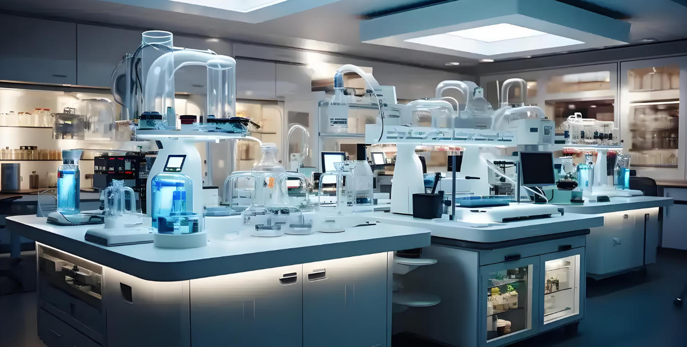
With the rapid advancement of life sciences, researchers increasingly require three-dimensional observation of cellular dynamics, tissue structures, and microscopic biological processes. However, traditional microscopy has clear limitations in real-time 3D imaging and high-precision data acquisition.
Time-of-Flight (ToF) microscopy, based on real-time depth sensing and high-speed 3D imaging, has emerged as a transformative tool for biological experiments. By leveraging its non-contact measurement and millisecond-level imaging speed, ToF microscopy enables high-precision visualization and intelligent data analysis, driving biological research toward greater accuracy, automation, and quantitative insight.
What Is Electron Microscopy Used For?
In biological research, electron microscopy (EM) remains an essential technique for nanoscale imaging and structure analysis. It is primarily used for the following:
High-Resolution Structural Observation
Electron microscopes use focused electron beams to capture nanoscale structures of cells, tissues, and biomaterials, revealing details far beyond the resolution of traditional optical microscopy.
Subcellular and Microscopic Tissue Analysis
In studies such as cell migration, neuronal outgrowth, and angiogenesis, EM provides highly detailed images of organelles, membranes, and tissue microstructures, forming a reliable foundation for quantitative biological data.
Multi-Modal Experiment Integration
Modern laboratories often integrate ToF microscopy and electron microscopy to combine advantages: ToF delivers real-time 3D spatial information, while EM provides ultra-high-resolution nanostructural data. This dual approach enables precise monitoring in cell motility, tissue engineering, and microscopic drug screening.
Data Visualization and Quantitative Analysis
By integrating ToF 3D point clouds and AI algorithms with EM images, researchers can perform quantitative morphology analysis, measuring cell volume, spatial distribution, and tissue organization for high-precision experimental evaluation.
In summary, electron microscopy is ideal for high-resolution structure observation and micro-level analysis, while ToF microscopy complements it with dynamic 3D imaging, making the two technologies highly synergistic for advanced biological research.
The Need for 3D Observation in Biological Experiments
In cutting-edge disciplines such as cell biology, tissue engineering, and microscopic drug discovery, researchers increasingly focus on 3D dynamic behaviors of cells and tissues. For instance:
In cell cultures, cells migrate, divide, and aggregate, forming complex 3D spatial networks.

In tissue engineering, cells grow within 3D scaffolds, reconstructing tissue architectures that mimic in vivo conditions.
In drug screening, drug responses evolve spatially and temporally within cellular or tissue environments.
Traditional 2D microscopy cannot capture these 3D processes accurately. Flat projections fail to represent cell migration trajectories, tissue spatial relationships, or microstructural transformations. For example:
In tumor metastasis studies, 2D images cannot quantify migration speed or direction in 3D space.
In neuroscience, 2D imaging cannot reconstruct complex 3D neuronal branching.
In angiogenesis experiments, 2D views cannot precisely measure vascular topology or branch angles.
Additionally, 2D observation often introduces error and bias. Researchers rely on multi-angle imaging or serial slicing to infer 3D structures—methods that increase complexity and reduce reproducibility, especially in live-cell dynamic imaging and high-throughput assays.
Therefore, real-time 3D dynamic observation has become essential in modern biological research. ToF microscopy fulfills this need by measuring the flight time of light pulses to reconstruct depth information, providing non-invasive, real-time 3D imaging at the microscopic scale. It enables accurate quantification of cell migration paths, tissue formation, and micro-drug effects, improving both reproducibility and scientific value.
In short, ToF microscopy establishes a new technological foundation for intelligent, high-throughput, and quantifiable 3D biological research.
Principles of ToF Microscopy
Time-of-Flight (ToF) microscopy is a high-precision 3D imaging technique that measures the travel time of light pulses to determine depth. Short infrared pulses are emitted toward a microscopic sample, and the time delay between emission and reflection is used to calculate spatial distances, generating comprehensive 3D depth maps.
1. Technical Advantages
3D Depth Perception
ToF microscopy provides complete 3D spatial data of samples, revealing cell morphology, layered tissue structures, and dynamic microenvironment changes—critical for cell migration analysis and tissue scaffold monitoring.
High-Speed, Real-Time Imaging
With millisecond-level acquisition rates, ToF captures rapid biological processes such as membrane oscillation, neural extension, angiogenesis, and cell proliferation, providing continuous 3D data streams.
Non-Contact Measurement
ToF imaging requires no staining or physical contact, avoiding interference with live cells, sensitive tissues, and delicate microstructures, preserving sample integrity while ensuring reliable data.
2. Comparison: ToF vs. Electron Microscopy (EM)
While electron microscopy achieves nanoscale resolution via electron beams, it requires extensive sample preparation (fixation, dehydration, coating) and cannot capture live or dynamic processes.
In contrast, ToF microscopy—though lower in resolution—offers real-time 3D observation, non-contact imaging, and live-sample compatibility, making it indispensable for continuous biological monitoring and dynamic process studies.

3. Applications in Biological Research
3D Cell Migration Tracking: ToF captures cell movement trajectories, speed, and shape changes in real time, ideal for cancer, immunology, and stem cell research.
Tissue Engineering: Enables live monitoring of cell distribution and tissue growth within 3D scaffolds.
High-Throughput Drug Screening: Allows non-contact evaluation of drug effects on cell behavior and proliferation.
Quantitative 3D Analysis: Combined with AI-driven image processing, ToF depth data supports automated volume and morphology measurement.
Live Environment Studies: Perfect for label-free, non-invasive 3D imaging of sensitive biological environments.
Experimental Cases and Achievements
1. Cell Migration Analysis
ToF microscopy enables real-time 3D tracking of individual or collective cell motion. In oncology, it quantifies cancer cell invasion through extracellular matrices; in immunology, it captures immune cell deformation and movement, providing valuable data for mechanistic studies.
2. Tissue Engineering and 3D Scaffold Observation
ToF microscopy monitors cell growth, distribution, and tissue remodeling inside scaffolds for organoid development—such as liver or cardiac tissue models—supporting data-driven quality control and optimization.
3. Data Visualization and Quantitative Analysis
High-precision ToF point clouds, combined with machine learning, allow automated measurement of cell volumes, morphology, and tissue formation rates, supporting AI-assisted biological imaging and reducing human error.
4. Live and Microenvironment Research
The non-contact ToF imaging method makes it ideal for live-cell studies, microfluidic systems, and drug response experiments, maintaining viability while continuously recording 3D dynamics.
Together, these use cases highlight ToF’s ability to deliver high-accuracy 3D data, real-time dynamics, and automated analysis, revolutionizing cellular biology, tissue engineering, and pharmaceutical screening.

Technical Challenges and Optimization Strategies
1. Challenges
Resolution and Sensitivity: Light scattering and attenuation at microscopic scales can reduce ToF accuracy for fine structures.
Dynamic Sample Motion: Cell movements can introduce instability in 3D imaging.
Environmental Interference: Ambient light or electromagnetic noise from lab equipment may affect signal quality.
Integration with Traditional Microscopy: Data fusion between ToF and EM remains technically complex.
2. Optimization Directions
Wavelength Optimization: Using near-infrared wavelengths enhances light penetration and reflection stability.
AI-Based Signal Processing: Deep learning algorithms enable noise reduction, multi-frame reconstruction, and real-time 3D modeling.
Multi-Modal Imaging: Combining ToF with EM and optical microscopes allows simultaneous dynamic 3D and nanoscale imaging.
Hardware Enhancements: Improved low-noise sensors, light source tuning, and expanded field of view increase stability and accuracy.
Dynamic Adaptation: Real-time depth calibration compensates for sample motion and environmental variation.
Through these advancements, ToF biological imaging can overcome its current limitations and deliver more precise, consistent, and efficient experimental outcomes.
Future Trends in ToF Microscopy for Life Sciences
1. ToF + AI for Automated 3D Experiments
Combining ToF microscopy with AI-based analytics enables:
Automated detection of cell migration and tissue growth patterns.
Precise 3D measurement of cellular structures.
High-throughput 3D drug screening with minimal human intervention.
2. Multi-Modal Fusion with Electron Microscopy
Integrating ToF with EM supports:
3D dynamic imaging plus ultra-high-resolution nanoscale analysis.
Cross-scale research from cell-level to subcellular-level structures.
Virtual experiment planning through 3D simulation prior to EM imaging.
3. Advanced Instrumentation
Next-generation ToF systems will feature:
Integrated sensors for temperature and humidity control.
Millisecond imaging rates for fast biological processes.
Safe, non-invasive operation ideal for live-cell research.
4. Toward Intelligent Laboratories
ToF microscopy combined with cloud-based AI platforms enables:
Remote experiment monitoring and control.
Automated report generation with quantitative 3D analysis.
Improved reproducibility and high-throughput experiment automation.
Conclusion
ToF microscopy—with its 3D observation, non-contact measurement, and high-speed imaging—is transforming biological research. From cell migration tracking to tissue engineering monitoring and high-throughput drug screening, ToF enhances experimental accuracy and streamlines workflows.
When integrated with AI analytics, multi-modal microscopy (such as EM), and intelligent lab systems, ToF will serve as a core enabling technology in next-generation life science laboratories—delivering more precise, efficient, and intelligent solutions for 3D biological imaging and analysis.
Synexens Industrial Outdoor 4m TOF Sensor Depth 3D Camera Rangefinder_CS40p

After-sales Support:
Our professional technical team specializing in 3D camera ranging is ready to assist you at any time. Whether you encounter any issues with your TOF camera after purchase or need clarification on TOF technology, feel free to contact us anytime. We are committed to providing high-quality technical after-sales service and user experience, ensuring your peace of mind in both shopping and using our products.
- このできごとのURL:


コメント